Is it bad for your health to eat food fried in olive oil?

DenisFilm/Shutterstock

James Brown, Aston University and Rachel Adams, Cardiff Metropolitan University

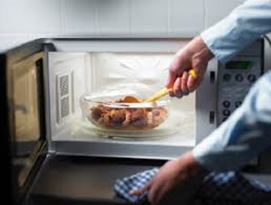
It recently has been suggested that using vegetable oils to fry food may be bad for your health due to the production of toxic chemicals called aldehydes during the heating process. Aldehydes are simple organic structures – compounds which contain a carbon-oxygen double bond – and are abundant in nature. They are formed in the human body in small amounts as by-products of normal fructose and alcohol metabolism. Consumption of dietary aldehydes is thought to contribute to human diseases including diabetes and heart disease. But what about olive oil? Is it classed as a vegetable oil, and is it safe to fry food with it?

Around half of UK households now use olive oil, which demonstrates a sizeable shift in our oil use in recent years. This may be in part because olive oil consumption is frequently linked to good health, and forms a central component of the Mediterranean diet. The Mediterranean diet is itself known to reduce the risk of disease and early death. Olive oil, produced by pressing olives, is commonly used across the world in food preparation, whether for frying, drizzling or as a part of a salad dressing. It is therefore classed as a vegetable oil, as it is produced from vegetable matter, as opposed to animal fats such as lard or goose fat.

Of the vegetable oils that have been tested for heating-induced aldehyde content, olive oil actually performs reasonably well. Researchers from the University of the Basque Country analysed olive, sunflower and flaxseed oils for their aldehyde content after the oils had been heated to 190℃. They found that heating the polyunsaturated sunflower and flaxseed oils produced greater quantities of aldehydes more quickly, whereas heating monounsaturated olive oil created fewer aldehydes and much later in the heating process.

This is thought to be because of a structural difference, with polyunsaturated oils containing more regions ripe for chemical reaction. Experiments performed for BBC’s Trust Me I’m a Doctor confirmed this, suggesting that heating olive oil, butter and goose fat produced similarly lower levels of aldehydes. These experiments collectively suggest that if you are going to fry, choosing olive oil is one of the better options.

Importantly, very little is known about what constitutes a low or high dose of aldehydes in food in humans. There is some data from animal studies, but the conclusions we can draw from them are limited. If olive oil is used to shallow fry foods for short periods, it is unlikely that your body would be exposed to greater concentrations of aldehydes than it normally would as a result of your body’s normal metabolic processes, mentioned earlier.

Verdict

While there are clearly healthier ways to cook foods, frying food with olive oil is unlikely to be significantly bad for your health.

Smoke points increase with olive oil quality.
Dusan Zidar/Shutterstock

Review

Rachel Adams, senior lecturer, Cardiff Metropolitan University

Heat causes chemical changes in all oils and this alters their aroma, flavour and nutritional content. Overheating oil during cooking will result in a dirty smoky kitchen, poor tasting food and the creation of harmful chemicals.

Olive oil is no different from other oils. If you burn it (heat it above its smoke point) it will taste bad and it will contain harmful chemicals. Smoke points tend to increase with olive oil quality, as the free fatty acid content tends to decrease and the antioxidant content increases. The high antioxidant content of olive oil could even reduce the amount of harmful chemicals produced during cooking. When cooking with olive oil, any potential harms can be reduced by using high quality oil and making sure you keep the oil below its smoke point; it will also make your food taste nicer.

The ConversationSo I agree with the author: frying in general is not the healthiest way to prepare food, but if you are going to fry then frying in olive oil is not a bad choice. If cooking with olive oil was that bad for you there would be population-based evidence to support the argument.

James Brown, Lecturer in Biology and Biomedical Science, Aston University and Rachel Adams, Senior Lecturer in Biomedical Science, Cardiff Metropolitan University

This article was originally published on The Conversation. Read the original article.

Is it safe to microwave your food?

Some say cancer-causing chemicals can leak into packaging and into your food.
osseous/Flickr, CC BY

Senaka Ranadheera, Victoria University; Duane Mellor, University of Canberra; Nenad Naumovski, University of Canberra, and Robyn McConchie, University of Sydney

Today every kitchen would seem “under-equipped” without a microwave, with its efficient ability to cook, defrost and reheat a variety of different foods. The handy appliance uses microwave radiation to do so. This is a type of electromagnetic radiation similar to radio waves and infrared light.

Although generally recognised as safe, the internet is awash with articles about the dangers microwave radiation poses to your food. Some claim using microwaves can cause “cataracts and cancer”. Other posts says it “zaps the nutrients right out of your food”.

If you believe this, the “killer” oven in your kitchen must be a terrifying sight, but there is actually no research to support the supposed dangers of microwave cooking. Hopefully we can allay your fears by checking some common danger claims against the evidence.

Does it zap the nutrients out?

Putting raw foods through any type of process – including heating and cooling – leads to changes in their physical properties, chemical composition and nutritional profile.

If nutrients are lost from foods cooked in microwaves, this would be because too high a temperature was used, or they were cooked for too long. The correct combination of time and temperature can help preserve most nutrients while also improving the foods’ taste, texture and colour.

The time and temperature required depends on the type of food. High risk foods such as meat, fish and eggs need to be heated to at least 60℃ to be safe.

Rapid cooking helps preserve beneficial chemicals in green vegetables.
from shutterstock.com

Microwave cooking is unlikely to negatively affect vitamins and other compounds associated with improved health. For instance, rapid cooking actually helps preserve a group of beneficial chemicals, the polyphenols – that increase the total antioxidant activity of foods – in green vegetables.

One study compared microwaving or steaming vegetables, such as cabbage, carrots, cauliflower and spinach, to pressure cooking. It found vegetables that were pressure cooked lost more insoluble fibre, which is good for gut health, than those that were microwaved or steamed.

A key nutrient usually destroyed when cooking vegetables is vitamin C, a severe lack of which can lead to conditions like scurvy. But boiling vegetables accounts for greater nutrient losses than microwaving them. This is because water soluble nutrients are readily leached into water when they are boiled, while very little water is used in microwaving.

Short bursts of heating, such as used in microwave cooking, can retain most of a vegetable’s vitamin C.

Can it give you cancer?

Some of the best studied cancer-causing compounds are the heterocyclic aromatic amines (HCA). These are formed naturally in protein-rich food such as meat and fish during cooking, and are more likely to form if the meat is cooked for a long time and at higher temperatures.

The method of cooking is a major factor affecting HCA formation. Some researchers have reported HCA are formed in chicken at higher levels when cooked in a microwave, compared to when pan-fried, barbecued or baked.

Barbecued fish has higher levels of HCA than microwaved fish.
from shutterstock.com

But no research has claimed or shown an association between regular consumption of microwave-cooked poultry and cancer.

A recent study has revealed barbecued fish contains more HCA than microwave-cooked fish, while HCA could not be detected at all in microwaved beef. Also, thawing beef and re-heating previously-cooked meat or fish in a microwave just for a few minutes, does not produce any extra HCA.

What about the packaging?

There is some evidence to suggest chemicals in plastic packaging can migrate into foods when microwaved, which has been associated with increased risk of cancer.

If your packaging has a microwave safe symbol, it is safe to use in the microwave.
from shutterstock.com

But most of today’s plastic containers, packages and wraps are specially designed to withstand microwave temperatures.

If packaging is marketed as microwave safe, has a microwave symbol or provides instructions for proper microwave use, it is safe for microwave cooking or heating.

Leaching of harmful toxins or “cancer-causing” compounds from appropriately packaged products during microwaving is highly unlikely in Australia, although this area could benefit from more research.

Does it kill bad bugs?

Cooking food significantly reduces the risk of food-borne illness.

A major challenge in microwaving is the unevenness of temperature distribution due to the shape of the food. You may notice when you heat food in a microwave that there are often hot and cold spots. This poses a potential safety issue.

Microwave cooking can only kill disease-causing bugs when the correct temperature and time combination is achieved throughout the food portion. Cooking to temperatures above 60℃ will kill most bugs known to cause food-borne illness, but the toxins produced by them may be heat-tolerant.

Stir food during the microwaving process so the heat is evenly distributed.
Lachlan Hardy/Flickr, CC BY

If the food is already contaminated with bugs that produce toxins, microwaving might kill the toxin-producing bug but not destroy the toxins, despite the correct temperature and time combination. This can also apply to other cooking methods. Appropriate food storage is the key to minimising such risks.

Minimising risk

  • Avoid overcooking vegetables to minimise nutrient losses
  • Before microwaving, check the labelling on the package and follow the instructions
  • If the package is not marked as being microwave-safe, switch to a suitable microwave container
  • Rotate and stir foods during cooking to spread the temperature of heating equally and as such minimise potential for food-borne illness. Check the temperature of food before consumption
  • The ConversationRemember microwaving cannot magically make contaminated food safe. So if in doubt, throw it out.

Senaka Ranadheera, Early Career Research Fellow, Advanced Food Systems Research Unit, College of Health and Biomedicine, Victoria University; Duane Mellor, Associate Professor in Nutrition and Dietetics, University of Canberra; Nenad Naumovski, Asistant Professor in Food Science and Human Nutrition, University of Canberra, and Robyn McConchie, Professor, Faculty of Agriculture and Environment, University of Sydney

This article was originally published on The Conversation. Read the original article.

No, you don’t have to finish all your antibiotics

Recommended antibiotic courses are often arbitrary.
Katy/Flickr, CC BY-NC-SA

Lyn Gilbert, University of Sydney

Most people believe – and have been told by health professionals – that it’s essential to finish a course of antibiotics to prevent antibiotic resistance. But this advice is not only wrong, it could actually be harmful.

The idea that you have to take all the antibiotics you’re prescribed is based on the assumption that all the bacteria causing the infection have to be killed, so the surviving minority don’t become resistant. In fact, for most otherwise healthy people, significantly reducing, but not necessarily totally eliminating, the bacteria causing the infection allows the body’s natural defences to take over and mop up the remaining few.

Some important caveats

There are some special circumstances when it’s important to kill all the bacteria – when the patient’s normal defences are damaged for any reason, for instance, or when the infection is in a site that’s relatively inaccessible to antibiotics and the white blood cells that kill bacteria. This can be in the middle of an abscess or cavity filled with pus (as in tuberculosis infection), on a foreign body, such as a prosthetic heart valve, or in dead tissue that can’t be removed (as in osteomyelitis or infection of the bone).

Obviously, stopping antibiotics before a serious infection is cured will risk a relapse. That’s what happened to Albert Alexander, the London policeman who was one of the first people to be treated with penicillin by Howard Florey in 1941.

Alexander had a terrible infection that started with a scratch on his face. He developed abscesses all over his head and had already had an eye removed, but he was dying.

Within 24 hours of being given a small dose of penicillin, his fever fell, his appetite returned and the abscesses started to heal. But when the penicillin supply ran out after five days, the infection flared up again. Alexander died four weeks later.

We now know that severe staphylococcal infection with multiple abscesses, which is what Alexander had, is a type of infection that needs antibiotic treatment for weeks to prevent relapse. But there’s a lot we still don’t know about the best way to treat some types of infection. It has recently become clear that some of the conventions around antibiotic prescribing are neither based on evidence nor harmless.

Antibiotics are generally benign but they all cause allergies and other rare side effects in a small proportion of people. And there’s a universal effect that’s less well known – even a very short course will kill many of the friendly bacteria in the gut.

The effect lasts for weeks, and the longer the antibiotic course, the greater the risk that antibiotic-resistant bacteria will take their place and cause harm. What’s more, they can spread to other people and add to the pool of antibiotic resistance in the community.

They can do worse damage too. Antibiotic-resistant bacteria include Clostridium difficile, which can be carried harmlessly in the bowel until a course of antibiotics kills off its competition. This allows it to multiply and produce toxins, potentially causing life-threatening diarrhoea.

Antibiotics are generally benign but they all cause allergies and other rare side effects in a small proportion of people.
Melissa Bowman/Flickr, CC BY

This, in turn, increases the risk of the bug spreading to other people, especially in hospitals and nursing homes where serious outbreaks often occur. Again, the longer the antibiotic course, the greater the risk of antibiotic-associated diarrhoea.

The right dose

The rate of antibiotic resistance (in a community, a hospital or a whole country) is proportional to the total amount of antibiotics used. The relationship is complex but the dangerous increase in multidrug-resistant bacteria has led some experts to predict the “end of the antibiotic era”. This is the downside of 75 years of antibiotic therapy.

Antibiotics have saved countless millions of lives, but have been often misused because of the misguided belief that they are harmless.

The most important – but hardly novel – message for doctors is “don’t prescribe antibiotics unnecessarily, especially for colds and flu, which are nearly always viral”. Antibiotics simply don’t work in acute upper respiratory infections. We all know from experience that a cough will often last for around ten days and there’s not a lot we can do to change that.

The problem is that it’s not always obvious whether some illnesses are due to infection and whether they are bacterial – and so might need treatment – or viral. Tests might help, but the patient would have to wait for results. So the decision to treat is usually based on clinical judgement – often influenced by the patient’s anxiety and the doctor’s (in)tolerance of risk.

The challenge for doctors and patients is to weigh the risks and benefits of treatment. Unless there are compelling reasons to start immediately, we should wait for test results or to see how symptoms develop. Equally importantly, we should stop the treatment immediately if, in hindsight, the diagnosis was wrong or symptoms disappear quickly.

Some serious bacterial infections, of course, need urgent and quite prolonged treatment. How long depends on the type of infection, how serious it is, the patient’s underlying condition and response to treatment.

But recommended antibiotic courses are often arbitrary; they may reflect long-standing convention or be based on a manufacturer’s decision during an initial drug trial. Recent clinical trials show that even for some serious infections, shorter antibiotic courses can be as effective as conventional, longer ones.

The ConversationThe general rule is: the shorter the course, the lower the risk of side effects or resistance. More trials are needed to determine the shortest courses that can be recommended without increasing the risk of relapse. But ultimately, it will still depend on clinical judgement not arbitrary rules, conventions or package inserts.

Lyn Gilbert, Clinical Professor in Medicine and Infectious Diseases, University of Sydney

This article was originally published on The Conversation. Read the original article.

Human genome editing:We should all have a say

Human genome editing: We should all have a say

Controversial gene editing should not proceed without citizen input and societal consensus.
(Shutterstock)

Françoise Baylis, Dalhousie University

Shoukhrat Mitalipov, a reproductive biologist at Oregon Health and Science University, is nothing if not a pioneer. In 2007, his team published proof-of-principle research in primates showing it was possible to derive stem cells from cloned primate embryos. In 2013, his team was the first to create human embryonic stem cells by cloning. Now, in 2017, his team has reported safely and effectively modifying human embryos with the MYBPC3 mutation (which causes myocardial disease) using the gene editing technique CRISPR.

Mitalipov’s team is not the first to genetically modify human embryos. This was first accomplished in 2015 by a group of Chinese scientists led by Junjiu Huang. Mitalipov’s team, however, may be the first to demonstrate basic safety and efficacy using the CRISPR technique.

This has serious implications for the ethics debate on human germline modification which involves inserting, deleting or replacing the DNA of human sperm, eggs or embryos to change the genes of future children.

Ethically controversial

Those who support human embryo research will argue that Mitalipov’s research to alter human embryos is ethically acceptable because the embryos were not allowed to develop beyond 14 days (the widely accepted international limit on human embryo research) and because the modified embryos were not used to initiate a pregnancy. They will also point to the future potential benefit of correcting defective genes that cause inherited disease.

This research is ethically controversial, however, because it is a clear step on the path to making heritable modifications – genetic changes that can be passed down through subsequent generations.

Beyond safety and efficacy

Internationally, UNESCO has called for a ban on human germline gene editing. And the “Convention for the Protection of Human Rights and Dignity of the Human Being with regard to the Application of Biology and Medicine” – the Oviedo Convention – specifies that “an intervention seeking to modify the human genome may only be undertaken for preventive, diagnostic or therapeutic purposes and only if its aim is not to introduce any modification in the genome of any descendants.”

In a move away from the positions taken by UNESCO and included in the Oviedo Convention, in 2015 the 12-person Organizing Committee of the first International Summit on Human Gene Editing (of which I was a member) issued a statement endorsing basic and preclinical gene editing research involving human embryos.

The statement further stipulated, however, that: “It would be irresponsible to proceed with any clinical use of germline editing unless and until (i) the relevant safety and efficacy issues have been resolved, based on appropriate understanding and balancing of risks, potential benefits, and alternatives, and (ii) there is broad societal consensus about the appropriateness of the proposed application.”

Mitalipov’s research aims to address the first condition about safety and efficacy. But what of the second condition which effectively recognizes that the human genome belongs to all of us and that it is not for scientists or other elites to decree what should or should not happen to it?

Modification endorsed

Since the 2015 statement was issued, many individuals and groups have tried to set aside the recommendation calling for a broad societal consensus.

For example, in February 2017, the U.S. National Academy of Sciences and National Academy of Medicine published a report endorsing germline modification. It states unequivocally that “clinical trials using heritable germline genome editing should be permitted” provided the research is only for compelling reasons and under strict oversight limiting uses of the technology to specified criteria.

Seeds of change in Canada

In Canada, it is illegal to modify human germ cells. Altering “the genome of a cell of a human being or in vitro embryo such that the alteration is capable of being transmitted to descendants” is among the activities prohibited in the 2004 Assisted Human Reproduction Act.

Worried that “Canadian researchers may fall behind on the international scene” and that “restrictive research policies may lead to medical tourism,” the Canadian Institutes for Health Research (with input from the Canadian Stem Cell Network) has begun to plant the seeds of change.

In its Human Germline Gene Editing report, CIHR hints at the benefits of changing the legislation. It also suggests professional self-regulation and research funding guidelines could replace the current federal statutory prohibition.

Future of the species

With Mitalipov’s technological advances and increasing suggestions from researchers that heritable modifications to human embryos be permitted, it is essential that citizens be given opportunities to think through the ethical issues and to work towards broad societal consensus.

We are talking about nothing less than the future of the human species. No decisions about the modification of the germline should be made without broad societal consultation.

The ConversationNothing about us without us!

Françoise Baylis, Professor and Canada Research Chair in Bioethics and Philosophy, Dalhousie University

This article was originally published on The Conversation. Read the original article.

How 50 medical experts separated Kenyan conjoined twins in 23-hour surgery

Former conjoined twins, Blessing and Favour, after a successful surgery at Kenyatta National Hospital.
Kenyatta National Hospital

Joseph Kimani Wanjeri, University of Nairobi

The successful separation of two year old conjoined twins at Kenyatta National Hospital marked a medical milestone in Kenya. Other than South Africa, very few successful separations have been performed in sub-Saharan Africa. In Kenya a multi-disciplinary team of medical experts operated on the twins for 23 uninterrupted hours. The Conversation Africa’s health and medicine editor Joy Wanja Muraya spoke to Dr Joseph Wanjeri about the surgery and post recovery of the twins who are home.

Can you explain conjoined twins, and its prevalence?

Conjoined twins are two babies born physically connected to each other. The extent and site of their union varies from sharing a band of skin and underlying tissues to more complex varieties sharing vital organs.

Research shows that cases of conjoined twins are found in one of every 50,000 live births globally. Kenya does not have a central repository for such cases.

Conjoined twins are the result of the embryo cells that have not completely separated. Embryo cells develop when the egg (ovum) is fertilised by sperm. They multiply and differentiate to form different body organs and tissues. An alternative theory is that two separate embryos fuse together in the early development of the twins.

The exact cause of conjoined twins is unknown but it’s thought that genetic factors interacting with environmental ones may contribute. Another possibility is the medicines taken by the mother during pregnancy.

The twins when they were first admitted at Kenyatta National Hospital on 5th September 2014. Photo: Author Provided.

Conjoined twins are classified based on the place they are joined. The most common types of conjoined twins are:

Seperating conjoined twins can be difficult and can result in death. Complex cases can be inoperable and others may call for emergency surgery soon after birth if the life of the twins is threatened.

Separating twins that are conjoined at the lower back (sacropagus) has fewer complications and deaths. The Kenyan twins were conjoined in this way.

Expertise, careful preparation and team work is the perfect recipe for a successful outcome of the surgery.

How is diagnosis done?

Diagnosis can be done before birth using an ultrasound scan or through physical examination at birth. Advanced tests like the Computerised Tomography (CT) scans and Magnetic Resonance Imaging (MRI) give greater details.

In the case of the Kenyan twins, a set of female conjoined twins was referred to the Kenyatta National Hospital in Nairobi in September 2014 from an upcountry health facility soon after their delivery.

The twins shared a spinal cord, rectum, anus, some muscles, subcutaneous tissues and skin. Paediatric, neuro and plastic surgeons agreed that separation was feasible but it should wait until the twins were bigger to withstand the complex surgery.

Paediatricians, nurses and nutritionists took care of the twins until they were one year old when the planning for the separation began. The plastic surgery team recommended tissue expansion, a procedure to help with closure of the huge soft tissue defects anticipated after separation. But the mother refused to give her consent, and the procedure had to be stopped. She clearly adored the twins but was overwhelmed by the decisions that needed to be taken. The matter was referred to court and the judge ruled that separation was in the best interests of the children. Their mother gave consent and the preparations for surgery were resumed.

A University of Nairobi plastic surgery resident created a 3D model of the twin’s pelvis to map the surgery.

A three-dimensional (3D) impression of the twins conjoinment. Kenyatta National Hospital.

Can you explain the details of the planning and actual surgery.

A multi-disciplinary medical team made up of 50 experts drawn from various medical fields did a dry run a week before the actual operation.

When the time came for the operation in November 2016 two sets of the anaesthetic teams took about three hours to anaesthetise and stabilise the twins.

The paediatric surgeons began the separation of the various soft tissues on one side up to the spine. The neurosurgeons took over and split the spine, opened the shared dura,which is the tough outermost membrane enveloping the brain and spinal cord. They later separated the nerves before repairing the dura.

The paediatric surgeons completed their separation of the soft tissue and successfully placed the pair on separate beds for the first time. But one was left without an anus and rectum. This condition would be repaired in the recovery phase.

The closure of the huge soft tissue defects and wounds was done on each of the girls by plastic surgeons. Local flaps and skin grafts covered the wounds after which an opening from the large intestine – a colostomy – was done.

They were transferred to the intensive care unit for specialised monitoring for two weeks before transfer to the paediatric surgical ward.

Blessing and Favour face each other for the first time since birth. Photo: Kenyatta National Hospital.

The wounds took about three months to heal. One of the twins developed impaired movement of one of her ankle joints. But after occupational therapy she was able to walk with only a slight limp.

The twin’s growth and development will be monitored in the coming months and further reconstructive surgeries scheduled at the appropriate time.

Blessing and Favour were discharged from hospital on June 15, 2017.

What does this success mean for sub-Saharan Africa and what lessons can be learnt?

First, it shows that medical experts in sub-Saharan Africa are qualified and capable of performing complex surgeries in spite of the poor resources at their disposal.

But it also shows that a referral centre with a multidisciplinary team is best suited for this kind of complex surgery.

The ConversationAnd as far as the children are concerned, it shows that intense nursing care, nutritional support, advanced wound care techniques and close monitoring by paediatric specialists has been critical to their remarkable recovery.

Joseph Kimani Wanjeri, Lecturer – Dept. of Surgery, Speciality: Plastic & Reconstructive Surgery , University of Nairobi

This article was originally published on The Conversation. Read the original article.

An extra organ or body part is more common than you think

Society has long treated people with extra limbs as anatomical oddities. But having an extra body part or organ is surprisingly common and many people don’t know they have them.
Ddicksson/Wikimedia Commons, CC BY-NC-SA

Michelle Moscova, UNSW

Doctors thought they were operating on a malignant tumour when they set about removing an unusual oval lump on the right side of a 40-year-old woman’s body. What they recovered instead was a perfectly normal and fully functioning extra spleen.

Most of us only have one spleen, an organ involved in immune function and blood filtering. But accessory or extra spleens are quite common, appearing in more than one in ten people.

It is not unusual for people with extra organs to be completely unaware of their existence. Often they are discovered accidentally during diagnostic scans for unrelated conditions. While many of these extra organs are rare, others are far more common than many of us believe. Some need to be surgically removed and others can be left alone.

The extra spleen, described above, is an example of what doctors call supernumeracy, when the body has an extra organ, part or structure.

Supernumeracy in history

Supernumeracy has long fascinated us, with many obvious and peculiar examples throughout history.

Witch hunters in the 16th and 17th centuries often identified supposed witches by their third nipple, although these extra nipples were often mistaken for moles or birthmarks.

Myrtle Corbin, with her four legs, was a side-show star. But most examples of supernumeracy are more subtle or hidden.
Wikimedia Commons

Then there are the famous cases in the era of the Barnum and Bailey freak shows, which displayed truly extraordinary examples of supernumeracy. These included the sideshow stars Frank Lentini, the three-legged man, and Myrtle Corbin, the four-legged woman.

Their conditions were the result of being attached to partially formed parasitic twins (also known as an asymmetrical or unequal conjoined twins) that had not fully separated during development. Both went on to marry other people and have normal children.

More recently was the internationally celebrated case of the eight-limbed Indian girl Lakshmi Tatma, born in 2005, who had four arms and four legs. Some considered her to be a reincarnation of a Hindu goddess. A 72-hour operation eventually separated her from her parasitic twin.

What causes supernumeracy?

Supernumeracy is caused by errors in how the embryo develops. While some of these conditions can be genetic, most occur spontaneously and have no known cause.

To understand how this happens it helps to think of the development of an embryo into a human as being like a finely tuned orchestra following the directions of a strict conductor.

Every player in the orchestra needs to know when to start playing and when to stop, how fast the pace should be and what instrument needs to dominate in every part of the symphony. If percussion plays too fast or strings come in too soon, it can end in a disaster.

Errors in embryonic development led to Indian girl Lakshmi Tatma being born with eight limbs.

Likewise, when the embryo develops, structures that will eventually make up a human baby need to fold, move, fuse and disappear at exactly the right time. If one structure persists too long or appears too early, it may block the way for another structure migrating to a new position. If a structure duplicates or fails to fuse with its other half, it can end up forming an extra organ.

What is most remarkable about the process of embryonic development is that in the great majority of cases we produce perfectly formed children.

This child developed extra toes as a result of exposure to the drug thalidomide in the womb.
Otis Historical Archives, National Museum of Health and Medicine/flickr, CC BY

In many cases of supernumeracy we don’t know what disrupted the development of the embryo, although in some cases a mother’s exposure to certain drugs or chemicals during pregnancy may be the cause.

One of the best known examples is thalidomide, a drug prescribed to pregnant women in the 1950s and 1960s to treat morning sickness but caused some 10,000 children worldwide to be born with significant birth defects.

Although absent or short limbs were among the most common birth defects reported, some babies had extra toes.

The drug was commonly taken in the first trimester of pregnancy when morning sickness is more common and when, coincidentally, the embryo develops most rapidly.

Why supernumeracy matters today

The historical cases highlighted earlier are all examples of extreme supernumeracy. But most cases of supernumeracy are so inconspicuous they are found by chance and have little impact on people’s lives.

Many people have extra teeth, like this 25-year-old man.
Dr Dalia Ibrahim/Radiopaedia.org, rID: 46513

In fact, most of the supernumerary organs we see in cadavers in the anatomy laboratory belong to body donors who were unaware of them during their lifetime.

Some supernumerary structures are very rare, including additional kidneys, penises or vaginas. Others are relatively common, including extra nipples and teeth.

Understanding these less obvious cases can be very important when diagnosing and treating patients. That’s why medical students learn about them.

One of the supernumerate structures taught in medical school is the cervical rib, an extra rib at the base of the neck above the normal rib cage, which occurs in about one in 200 people.

While not apparent without diagnostic imaging, it can compress nerves and blood vessels that pass between the neck and shoulder, leading to numbness and pain in the arms and fingers. So, every medical student is taught about this anatomical variation.

Being aware of supernumerate structures can help doctors make a correct diagnosis.
For example, about one in 2,000 of us have an extra ureter – a muscular tube that carries urine from the kidneys to the bladder, where it is stored until ready to be excreted through the urethra. While an extra ureter does not necessarily cause problems, in some people the ureter connects the kidney with the wrong structure, for example with the vagina or urethra.

Extra nipples are another common example of supernumeracy.
Zureks/Wikimedia Commons, CC BY-SA

This means the ureter bypasses the normal mechanism that stops urine from leaking out of the bladder. This anomaly is usually noticed in childhood, as patients who have this type of ureter often have continuous dripping of urine which needs to be surgically corrected.

Doctors also need to be aware that for patients with supernumerary organs, these organs need to be included in cancer screening.

For example, if a patient has supernumerary breasts, these breasts need to be included when screening for lumps or in mammograms.

Even the smallest supernumerary structure may alter some body functions, as it did for a 29-year-old optometry student who in a practical class accidentally discovered she had an extra opening in her eyelid known as a punctum.

The punctum helps drain away excessive tears. Usually, we only have one in each eyelid but this student had two in the same eyelid.

This explained why anaesthetic eye drops used for optometry procedures were far less effective in that eye, as the drops drained away twice as fast.

Knowing that she had a supernumerary punctum, doctors told her to press down on it to close off the opening when she needed to use eye drops.

The ConversationSo, while many of us may be aware of supernumerary structures because of the extraordinary examples usually related to parasitic twins, the more subtle supernumerary structures, which are far more common, have an important place in the study of anatomy and medical practice.

Michelle Moscova, Senior Lecturer in Anatomy, UNSW

This article was originally published on The Conversation. Read the original article.

Are maternal hormones different when carrying a boy or a girl?

Will a female pregnancy make mum more emotional, and a male one more aggressive?
from www.shutterstock.com.au

Monique Robinson, University of Western Australia

With advances in prenatal testing it’s now possible to find out whether a pregnancy will result in a male or female baby as early as eight weeks’ gestation.

A quick check of web pregnancy forums would indicate that wait is too long for many parents-to-be who are excited to predict the sex of their baby by interpreting symptoms of pregnancy that might be related specifically to boys or girls.

The old wives’ tales and myths abound when it comes to predicting gender in pregnancy. For example, girls will steal your beauty, a boy will make your hair grow faster, girls will make you emotional, and boys will make you aggressive.

Did these myths grow from some kernel of truth that might be backed up by scientific evidence?

Hormone differences for baby boys and girls

There is evidence hormone concentrations in pregnancy can differ according to the sex of the fetus as early as three weeks after fertilisation. Studies have shown hCG (human chorionic gonadotropin, the hormone responsible for the second line appearing on a home pregnancy test) is higher for female fetuses compared with males, and remains higher throughout pregnancy. Some studies only report this later in pregnancy.

The surge of hCG early in pregnancy is one explanation for the less desirable symptoms of pregnancy, such as nausea and sickness. Researchers have found that severe morning sickness (called hyperemesis gravidarum) is slightly more common in pregnancies where the baby is a girl, which could reflect the differences in hCG.

There are plenty of other hormones at work in pregnancy, whatever the sex of the fetus. There’s a lay belief that when pregnant with a girl, maternal oestrogen levels are higher, but this is not backed up by most studies. Actually, maternal blood oestrogen levels rise steadily throughout pregnancy whatever the sex of the fetus, although female fetuses do show a higher oestrogen concentration in the amniotic fluid early in the second trimester.

Testosterone in the maternal bloodstream follows a similar pattern, increasing slowly throughout pregnancy with no fetal gender-related differences. Once again, within the amniotic fluid there are differing concentrations of testosterone though, higher for males than females.

Evidence doesn’t support higher levels of the male or female hormone in the mother’s bloodstream depending on the sex of the fetus.
from www.shutterstock.com

Differences in maternal oestrogen levels in maternal blood have been reported though. In one Scandinavian study, oestrogen levels in the first half of pregnancy were around 9% higher for pregnancies where the baby was female, and progesterone levels were lower in the second trimester.

But the extent to which the maternal hormone levels matches up to the fetal hormones is difficult to predict. The fetus is influenced by its own internal hormones in addition to being exposed to circulating maternal hormones. Getting samples of fetal blood is complicated and risky, so there is much we don’t know about this relationship.

There is great interest in this type of research beyond parents who’d like to get the nursery painted early. The field of the developmental origins of health and disease focuses on how the environment in the uterus, including the hormonal environment, might affect later health, behaviour and well-being.

Hormonal imbalances in the uterus have been linked with heart disease, cancer, polycystic ovary syndrome and autistic behaviours in the offspring.

Are pregnancies with girls and boys different?

If there was an oestrogen surge in mothers who are expecting a female baby, could this explain some of the old wives’ tales of extra moodiness or teariness in girl pregnancies?

There are significant individual differences in how vulnerable women are to fluctuations in hormones. In some cases, higher oestrogen levels have been reported as “psycho-protective”. This means they moderate mood, while other authors report oestrogen fluctuations triggering mood disorders and even depression in women.

What about aversions or nausea that women report to be different in male and female pregnancies?

A fascinating 2015 study looked at the emotion of disgust and how it differs according to the sex of the baby. The researchers posited that disgust is a protective mechanism that helps the mother avoid risks and harms during pregnancy – for example, aversions to certain foods or situations.

They found that disgust was high in the first trimester for pregnancies bearing boys and girls, but it decreased for girl pregnancies after the first trimester and remained high for boy pregnancies into the second trimester.

The reasoning for this is that male fetuses are considered more vulnerable to their environment, and the aversions reflect a natural predisposition to maintain protective behaviours longer for male pregnancies than female pregnancies.

The ConversationSo it appears there’s not enough evidence to back up claims male or female pregnancies differ significantly in terms of the maternal hormonal environment. This makes it unlikely that anecdotes of moodier, angrier or uglier pregnancies are due to the sex of the fetus.

Monique Robinson, Associate Principal Investigator, Telethon Institute for Child Health Research, University of Western Australia

This article was originally published on The Conversation. Read the original article.