Seven body organs you can live without

Komsan Loonprom/Shutterstock

Adam Taylor, Lancaster University

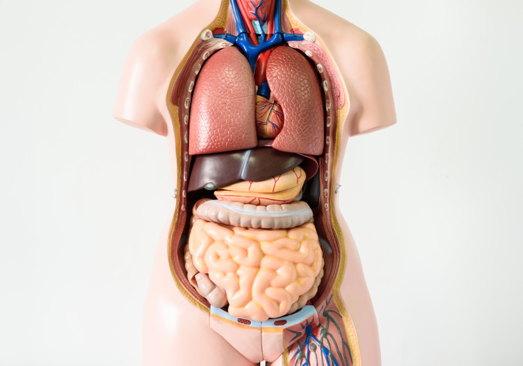
The human body is incredibly resilient. When you donate a pint of blood, you lose about 3.5 trillion red blood cells, but your body quickly replaces them. You can even lose large chunks of vital organs and live. For example, people can live relatively normal lives with just half a brain). Other organs can be removed in their entirety without having too much impact on your life. Here are some of the “non-vital organs”.

Spleen

This organ sits on the left side of the abdomen, towards the back under the ribs. It is most commonly removed as a result of injury. Because it sits close the ribs, it is vulnerable to abdominal trauma. It is enclosed by a tissue paper-like capsule, which easily tears, allowing blood to leak from the damaged spleen. If not diagnosed and treated, it will result in death.

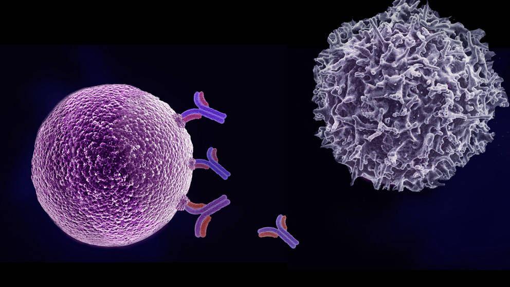
When you look inside the spleen, it has two notable colours. A dark red colour and small pockets of white. These link to the functions. The red is involved in storing and recycling red blood cells, while the white is linked to storage of white cells and platelets.

You can comfortably live without a spleen. This is because the liver plays a role in recycling red blood cells and their components. Similarly, other lymphoid tissues in the body help with the immune function of the spleen.

Stomach

The stomach performs four main functions: mechanical digestion by contracting to smash up food, chemical digestion by releasing acid to help chemically break up food, and then absorption and secretion. The stomach is sometimes surgically removed as a result of cancer or trauma. In 2012, a British woman had to have her stomach removed after ingesting a cocktail that contained liquid nitrogen.

When the stomach is removed, surgeons attach the oesophagus (gullet) directly to the small intestines. With a good recovery, people can eat a normal diet alongside vitamin supplements.

Just say no.
Nazar Skladanyi/Shutterstock

Reproductive organs

The primary reproductive organs in the male and female are the testes and ovaries, respectively. These structures are paired and people can still have children with only one functioning.

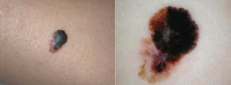
The removal of one or both are usually the result of cancer, or in males, trauma, often as a result of violence, sports or road traffic accidents. In females, the uterus (womb) may also be removed. This procedure (hysterectomy) stops women from having children and also halts the menstrual cycle in pre-menopausal women. Research suggests that women who have their ovaries removed do not have a reduced life expectancy. Interestingly, in some male populations, removal of both testicles may lead to an increase in life expectancy.

Colon

The colon (or large intestine) is a tube that is about six-feet in length and has four named parts: ascending, transverse, descending and sigmoid. The primary functions are to resorb water and prepare faeces by compacting it together. The presence of cancer or other diseases can result in the need to remove some or all of the colon. Most people recover well after this surgery, although they notice a change in bowel habits. A diet of soft foods is initially recommended to aid the healing process.

Gallbladder

The gallbladder sits under the liver on the upper-right side of the abdomen, just under the ribs. It stores something called bile. Bile is constantly produced by the liver to help break down fats, but when not needed in digestion, it is stored in the gallbladder.

Gallstones.
Martin Charles Hatch/Shutterstock

When the intestines detect fats, a hormone is released causing the gallbladder to contract, forcing bile into the intestines to help digest fat. However, excess cholesterol in bile can form gallstones, which can block the tiny pipes that move bile around. When this happens, people may need their gallbladder removed. The surgery is known as (cholecystectomy. Every year, about 70,000 people have this procedure in the UK.

Many people have gallstones that don’t cause any symptoms, others are not so fortunate. In 2015, an Indian woman had 12,000 gallstones removed – a world record.

Appendix

The appendix is a small blind-ended worm-like structure at the junction of the large and the small bowel. Initially thought to be vestigial, it is now believed to be involved in being a “safe-house” for the good bacteria of the bowel, enabling them to repopulate when needed.

Due to the blind-ended nature of the appendix, when intestinal contents enter it, it can be difficult for them to escape and so it becomes inflamed. This is called appendicitis. In severe cases, the appendix needs to be surgically removed.

A word of warning though: just because you’ve had your appendix out, doesn’t mean it can’t come back and cause you pain again. There are some cases where the stump of the appendix might not be fully removed, and this can become inflamed again, causing “stumpitis”. People who have had their appendix removed notice no difference to their life.

Kidneys

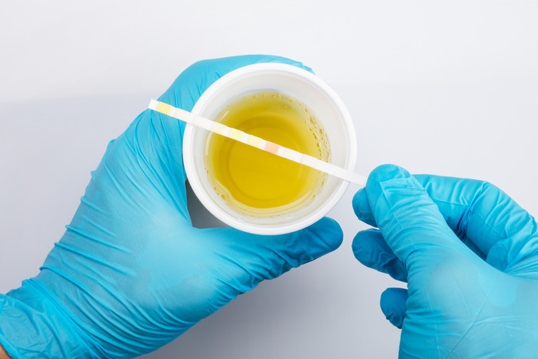
Most people have two kidneys, but you can survive with just one – or even none (with the aid of dialysis). The role of the kidneys is to filter the blood to maintain water and electrolyte balance, as well as the acid-base balance. It does this by acting like a sieve, using a variety of processes to hold onto the useful things, such as proteins, cells and nutrients that the body needs. More importantly, it gets rid of many things we don’t need, letting them pass through the sieve to leave the kidneys as urine.

There are many reasons people have to have a kidney – or both kidneys – removed: inherited conditions, damage from drugs and alcohol, or even infection. If a person has both kidneys fail, they are placed onto dialysis. This comes in two forms: haemodialysis and peritoneal dialysis. The first uses a machine containing dextrose solution to clean the blood, the other uses a special catheter inserted into the abdomen to allow dextrose solution to be passed in and out manually. Both methods draw waste out of the body.

The ConversationIf a person is placed on dialysis, their life expectancy depends on many things, including the type of dialysis, sex, other diseases the person may have and their age. Recent research has shown someone placed on dialysis at age 20 can expect to live for 16-18 years, whereas someone in their 60s may only live for five years.

Adam Taylor, Director of the Clinical Anatomy Learning Centre & Senior Lecturer in Anatomy, Lancaster University

This article was originally published on The Conversation. Read the original article.

Spot the difference: harmless mole or potential skin cancer?

If you’re at high risk of skin cancer, check your skin regularly.
Roman Königshofer/Flickr, CC BY-ND

H. Peter Soyer, The University of Queensland and Anna Finnane, The University of Queensland

The earlier you find a cancerous mole, the easier the treatment and the better the outcomes. But it’s not easy distinguishing between harmless, benign moles and those that warrant further attention.

In recent decades, the incidence of skin cancer has increased in Australia. Two in three Australians will be diagnosed with skin cancer by the time they are 70 years old.

Non-melanoma skin cancers, including basal cell carcinoma and squamous cell carcinoma, are the most common skin cancers but are less dangerous than melanoma.

In 2010, melanoma was the fourth most commonly diagnosed cancer in Australia, with 11,405 new cases diagnosed. It is also the most common cancer diagnosed in Australians aged 15 to 39 years.

A number of characteristics are associated with an increased risk of melanoma, including:

  • age
  • number of moles
  • skin type and colour (especially if you always burn and never tan in the sun)
  • personal history of melanoma or other skin cancer
  • freckles
  • unusual-looking moles, larger than five millimetres
  • red or light hair.

High levels of sun exposure and history of sunburn also increase the risk of melanoma.

Advances in treatment over the past three decades have improved the chances of survival. The five-year survival rate has increased from 85.8% to 90.7%. The prognosis is better the earlier melanoma is diagnosed.

Bad sunburn increases your risk of melanoma.
Kevin O’Mara/Flickr, CC BY-NC-ND

By regularly checking your own skin, you may notice moles that are changing as well as identify new moles. A study of more than 3,500 Queenslanders with melanoma found that almost half of the melanomas were detected by patients themselves and a fifth were found by partners.

The features of melanoma to look out for are often referred to as the ABCD rule:

  • Asymmetry
  • Border irregularity
  • Colour variation
  • Diameter larger than five millimetres.

Doctors and the wider population have used this rule for more than 25 years to identify suspicious moles. But with the increasing diagnosis of smaller melanomas and border irregularity being a feature of many benign moles, these features are not always good predictors of whether a mole is cancerous or not.

In 2011, a simpler AC (Asymmetry and Colour) rule was proposed for wider use by the community. While normal moles tend to be symmetrical and uniform in colour, melanomas tend to be asymmetrical and display multiple colours.

Asymmetry and colour variation typical of melanoma.
Peter Soyer

Using this rule, people without any professional health care experience were able to correctly identify 93% of melanoma images.

Symmetry and consistent colour of benign moles.
Peter Soyer
Peter Soyer

Not all melanomas follow these rules and some are very difficult to identify. In particular, a type of melanoma called nodular melanoma is a fast-growing, aggressive form of melanoma, which has poorer treatment outcomes and survival.

Researchers in Melbourne proposed the addition of “EFG” (elevated, firm and growing for more than a month) to the widely known ABCD rule, to improve early detection of nodular melanoma. These melanomas often begin as a red nodule. While their appearance can be mistaken for a pimple, they are much firmer to touch. This should prompt further investigation.

Nodular melanomas are fast-growing and aggressive.
Peter Soyer

If you’re concerned about moles with any of these features, consult your GP, who can refer you to a dermatologist if warranted.

If you’re at high risk, based on the risk factors listed above, check your skin regularly. Have another person (partner, family member or GP) check hard-to-see areas such as the back, neck and scalp.

Better melanoma detection

As awareness of melanoma has increased and consumers have taken a more active role in their own health care, the technological world has responded with new hardware to transform smartphones into diagnostic medical instruments and software designed to “autodiagnose” skin cancers.

Astute entrepreneurs have developed numerous mobile applications aimed not only at educating and calculating individual risk, but also enabling people to document, track and analyse their own moles.

But experts have raised questions about their accuracy. A recent study submitted images of 15 melanocytic lesions (five melanomas and ten benign moles) to five different mobile applications, to compare the risk grade allocated by each app (low, moderate or high risk for melanoma) with the clinical diagnosis.

Two of the five apps tested did not identify any of the melanomas. The other three apps detected 80% of the melanomas.

While the technology is exciting and it can be tempting to engage in this new mode of health care, be wary, particularly about applications costing money. The accuracy of many of these apps is questionable and could give false reassurance and lead to delayed diagnosis.

Research is continuing into combining the use of mobile dermatoscopes, a device using a light source and magnifying lens that can be attached to a smartphone to examine the skin, along with specialist advice.

Mobile teledermoscopes attach a light source and magnifying lens to a smartphone.
Queensland University of Technology

Using such a device, people can take images of their own moles and store them to monitor changes in moles over time, as well as send them to a dermatologist for diagnosis. This process is called teledermoscopy.

So far, studies suggest consumers are able to identify suspicious moles and take high-quality images that are adequate for a specialist diagnosis. But specialists are concerned that consumers may overlook moles that would have been worthwhile photographing.

Researchers are now investigating the factors influencing wider application in the health-care setting. The benefits of consumer-led mobile teledermoscopy could include reduced waiting times, reduced costs, earlier diagnosis and improved treatment outcomes.

The ConversationMobile dermatoscopes are likely to become a popular smartphone accessory in the future. Until then, the best we can do is regularly check our skin using the ABCD/AC and EFG rules, and show any suspicious mole to a GP or dermatologist.

H. Peter Soyer, Professor of Dermatology, The University of Queensland and Anna Finnane, Post-doctoral Research Fellow, The University of Queensland

This article was originally published on The Conversation. Read the original article.

Who lives longest: meat eaters or vegetarians?

Lukasz Szwaj/Shutterstock.com

James Brown, Aston University

Our ability to live a long life is influenced by a combination of our genes and our environment. In studies that involve identical twins, scientists have estimated that no more than 30% of this influence comes from our genes, meaning that the largest group of factors that control how long a person lives is their environment.

Of the many possible environmental factors, few have been as thoroughly studied or debated as our diet. Calorie restriction, for example, is one area that is being investigated. So far, studies seem to show that restricting calories can increase lifespan, at least in small creatures. But what works for mice doesn’t necessarily work for humans.

What we eat – as opposed to how much we eat – is also a hot topic to study and meat consumption is often put under the microscope. A study that tracked almost 100,000 Americans for five years found that non-meat eaters were less likely to die – of any cause – during the study period than meat eaters. This effect was especially noticeable in males.

Some meta-analyses, which combine and re-analyse data from several studies, have also shown that a diet low in meat is associated with greater longevity and that the longer a person sticks to a meat-free diet, the greater the benefit. Not all studies agree, however. Some show very little or even no difference at all in longevity between meat eaters and non-meat eaters.

What is clear is evidence that meat-free diets can reduce the risk of developing health problems such as type 2 diabetes, high blood pressure and even cancer. There is some evidence to suggest that vegan diets possibly offer added protection above a standard vegetarian diet. These findings are far easier to interpret as they report the actual event of being diagnosed with a health problem rather than death from any cause.

So can we confidently say that avoiding meat will increase your lifespan? The simple answer is: not yet.

The problem with longevity

The first thing that is clear is that, compared with most other creatures, humans live for a very long time. This makes it very difficult to run studies that measure the effect of anything on longevity (you’d have difficulty finding a scientist willing to wait 90 years for a study to complete). Instead scientists either look back at existing health records or recruit volunteers for studies that use shorter time periods, measuring death rates and looking to see which group, on average, was mostly likely to die first. From this data, claims are made about the effect certain activities have on longevity, including avoiding meat.

There are problems with this approach. First, finding a link between two things – such as eating meat and an early death – doesn’t necessarily mean one thing caused the other. In other words: correlation does not equal causation. It may appear that vegetarianism and longevity are related but a different variable may explain the link. It could be that vegetarians exercise more, smoke less and drink less alcohol than their meat eating counterparts, for example.

Maybe vegetarians exercise more than meat eaters.
Rasulov/Shutterstock.com

Nutrition studies also rely on volunteers accurately and truthfully recording their food intake. But this can’t be taken for granted. Studies have shown that people tend to underreport calorie intake and overreport healthy food consumption. Without actually controlling the diet of groups of people and measuring how long they live, it is difficult to have absolute confidence in findings.

The ConversationSo should I avoid meat for a long and healthy life? The key to healthy ageing probably does lie in controlling our environment, including what we eat. From the available evidence it is possible that eating a meat-free diet can contribute to this, and that avoiding meat in your diet could certainly increase your chances of avoiding disease as you age. But there’s certainly also evidence to suggest that this really might work in tandem with avoiding some clearer risks to longevity including smoking.

James Brown, Lecturer in Biology and Biomedical Science, Aston University

This article was originally published on The Conversation. Read the original article.

Michael Douglas, oral sex and cancer – the facts about HPV

An estimated 5.2% of cancers worldwide are caused by the human papilloma virus.
Image from shutterstock.com

Dyani Lewis, University of Melbourne

Oral sex is often the first of many forays into sexual intimacy we experience as fumbling teenagers. And for many couples, it remains an important – and enjoyable – part of their sexual repertoire. But can it cause cancer?

Michael Douglas has bravely declared that cunnilingus was the cause of his throat cancer. In particular, Douglas pointed the finger at the human papilloma virus, or HPV.

What is HPV?

HPV is not a single virus, but a family of more than 120 known viruses that can infect our body’s surfaces – our skin and mucous membranes.

A large number of these viruses go completely unnoticed – our immune system effectively clears them without us ever knowing we were infected. Others cause common warts, flat warts, plantar warts, and genital warts. Any area of skin that gets a graze can potentially become infected.

And then there are the HPV types that cause cancer, the oncoviruses. Current estimates put the number of oncoviruses at around 20, although a handful of culprits account for the lion’s share of HPV-caused cancers: HPV16 and 18 cause around 70% of cervical cancers.

The human papilloma virus.
Image from shutterstock.com

In humans, HPV isn’t the only cancer-causing virus – the hepatitis viruses B and C can cause liver cancer, for example, and the Epstein–Barr virus causes a range of cancers including Hodgkin’s lymphoma.

But in terms of sheer numbers that it affects, HPV tops the list. In 2002, the World Health Organization estimated that 5.2% of all cancers worldwide were caused by HPV.

Most people who contract HPV – even a cancer-causing variety – will naturally clear the infection within a few months. But for an unlucky few, the infection persists for years, putting them at risk of developing cancer.

Cervical cancer

HPV16 and 18 have been known causes of cancer in humans since the early 1980s, when it was identified as a common culprit of cervical cancers in women.

By 1991, a cervical cancer screening program was established in Australia and the biennial visit to the GP for a Pap smear commenced for all women aged 18 to 69 years, as well as for younger women two years after first sexual intercourse.

As much as women might cringe at the thought of a Pap smear, regular screening is a crucial means of identifying the tell-tale anatomical changes that are the prelude to cervical cancer formation. For around 90% of cases, early intervention after a Pap smear – including removal of the offending tissue – is effective at preventing cancer from developing.

Cancers of the throat are increasingly being attributed to HPV infection.
Image from shutterstock.com

Throat cancers

It is only more recently, in 2004, that HPV infection was clearly linked with cancers of the head and neck. Just as the cervix at the top of the vagina is an attractive landing ground for HPV infection, so too is the surface of the tonsils and the back of the tongue. Together, these fleshy regions at the back of our mouth make up the oropharynx, a large part of what we commonly call the throat.

While cancers of the lips, gums, and front portion of the tongue are usually the result of smoking or chewing tobacco, or drinking alcohol, cancers of the throat are increasingly being attributed to HPV infection. HPV16 is again the primary culprit.

Smoking-related head and neck cancers have been falling in Australia and other developed countries that have vigorous anti-smoking campaigns. At the same time, oropharyngeal (throat) cancers due to HPV are on the rise. An Australian study found that in 1987, just one in five throat cancers tested positive for HPV16 or 18. In 2005/06, the proportion had leapt to two-thirds.

Cancer prevention

As with any cancer, prevention is better than cure. The HPV vaccine protects against the two main types of cancer-causing HPV (HPV16 and 18), as well as two types that cause genital warts.

Clinicians have already seen a dramatic decrease in genital warts in young women since the vaccination program was introduced in mid-2007; the prevalence of genital warts plummeted from 9.6% in 2004, to less than 2% in 2010-11.

It is likely that a similar decline in throat cancers will result from vaccination, especially as boys are included in the immunisation program in Australia.

Condoms and dental dams can help prevent transmission.
Flickr/lil latvian

One curious point that Douglas made with his announcement was that cunnilingus was the best cure for HPV infection.

Unfortunately there is no therapeutic benefit to cunnilingus. HPV is transmitted, along with many other sexually transmitted infections such as HIV, chlamydia and gonorrhoea, via both genital and oral contact.

Condoms and dental dams can go some distance to preventing transmission, but touching, kissing and anything else that transfers body fluid from one person to the next can transmit the infection.

The ConversationAside from safer sex, a regular trip to the dentist to catch cellular changes early and a jab with the HPV vaccine are the best ways of preventing HPV-related throat cancer.

Dyani Lewis, Sexual health researcher, University of Melbourne

This article was originally published on The Conversation. Read the original article.

BCAA supplements are just hype – here’s a better way to build muscles

So macho.
Aleksandra Gigowska

Lee Hamilton, University of Stirling; Brad Schoenfeld, City University of New York, and Kevin Tipton, University of Stirling

As gym regulars we never cease to be amazed at the array of post-workout concoctions people consume in the changing room. We see everything from pills and powders to a rainbow of luminous drinks. Mostly it’s with one goal in mind, of course: to obtain muscles as close to the models endorsing these supplements as you possibly can.

The global market for sports nutrition products is predicted to hit $45 billion (£33 billion) by 2022, an increase of about 60% on 2016 value. Previously the domain of bodybuilders and elite athletes, amateur exercisers are becoming big supplement consumers thanks to the rising popularity of obstacle races, boot camps and CrossFit – all of which the product manufacturers target heavily.

One particularly popular variety is products containing branched-chain amino acids (BCAAs). These seem to promise all the benefits of boosting muscle-building after exercise with none of the hassle of foods. But do these products work – or are there better ways to help you get the most out of your gym membership?

Food vs supplements

The protein in our muscles is constantly turning over, simultaneously being broken down and built up (“synthesised”). In young healthy adults, the rate at which these two processes occur is usually the same. But to grow muscles, the rate of synthesis has to be greater than the rate of breakdown. You can do this by combining a workout of lifting weights with consuming either a foodstuff rich in protein, such as whole milk, or the isolated amino acids derived from protein.

Block-headed.
PowerUp

Amino acids are the building blocks of protein and muscle. They come in 22 different varieties. A range of experiments has shown that three of these are particularly effective at activating the machinery responsible for increasing muscle protein synthesis. They are leucine, isoleucine and valine – collectively known as the BCAAs.

Supplement companies sell many products that contain just BCAAs, based on various findings about their importance, which mostly come from animal tests. Indeed, our own work suggests that leucine is particularly potent for activating the body’s protein synthesis machinery. Many people take this to mean that consuming isolated BCAAs will therefore generate a growth response as great as from an equivalent amount of food, without the additional calories or logistical problems of taking a meal to the gym.

Yet recent work from our respective research teams suggests otherwise. We know for example that you can achieve a substantial increase in protein synthesis from 20g of whole egg protein, which is around three large eggs, or from 20g of whey protein. (Though to get the maximum possible response after a full body workout, our research on whey suggests it may be necessary to consume as much as 40g.)

Under very similar lab conditions to the latter whey study, we gave a similar group of male subjects 5.6g of BCAAs following a session of lifting weights – the equivalent to 20g of whey. The resulting protein synthesis response was only about 22% – or about half what would be achieved with the equivalent dose of whey.

Whey to go.
Jasminko Ibrakovich

What would the reason for this be? Various studies suggest that to achieve a full growth response, you need a sufficient supply of the whole complement of amino acids. This seems logical: think of it like building a brick wall where 20 different bricks are required to complete the wall. The BCAAs might act like the site foreman in bringing together all the men and machinery required to build the wall. But you still can’t build the wall without the rest of the bricks, no matter how much the foreman berates the bricklayers.

In other words, following exercise you need whole protein sources that provide a high dose of BCAAs and sufficient amounts of all the other amino acids – for example meat, dairy or eggs. And not only are BCAAs less equipped to build the muscle wall, consuming them on their own potentially creates a competition with other amino acids for absorption through the intestinal wall. If so, they could be making it harder for the body to gather up the other “bricks” essential to muscle building.

Window dressing

Besides building muscle, people also consume BCAAs (and other supplements) to allow them to hit the so-called anabolic window – the period of time available post-workout, often said to be between 30 minutes and two hours, in which to consume protein/amino acids to get the benefits of muscle building.

Our work indicates that the timing of post-workout nutrition is not as important for building muscle as once thought, however. There is time to get home and cook a meal, possibly even as much as 48 hours, so long as it contains between 25g and 30g of high-quality protein. To maximise muscle building you should also ensure you get around 1.6g of protein per day for each kilogram of your weight (that’s about 130g for an 80kg person).

Finally, there are studies which suggest potential benefits of BCAAs when it comes to optimising body composition – maintaining muscle mass at the same time as dropping fat. Yet these have been criticised for improper statistical analysis and inconsistencies in reporting of data.

Protein power.
Oleksandra Naumenko

So where muscle building is the goal, we support the “food first” approach recommended by the Sports and Exercise Nutrition Register. This means sticking with foods unless you’re struggling to find time to hit the necessary protein levels – in which case, a quick whey shake may be a convenient and effective alternative.

Consuming protein-rich foods also contributes to the daily nutritional needs for a range of minerals and vitamins – something often lacking in supplements. Sticking with food will also help you avoid the risk of consuming a “banned” substance through supplementation. One study from 2007 tested 58 supplements and found that about a quarter were contaminated with banned substances.

The ConversationIn short, don’t be taken in by the expensive luminous drinks in the changing rooms. Whatever it may say on the side of the bottle, they make muscle-building much more complicated and inefficient than it ever has to be.

Lee Hamilton, Lecturer in Sport, Health and Exercise Science, University of Stirling; Brad Schoenfeld, Assistant Professor, Exercise Science, City University of New York, and Kevin Tipton, Professor of Sport, Health and Exercise Sciences, University of Stirling

This article was originally published on The Conversation. Read the original article.

Buruli ulcer: Africa’s neglected but third most common mycobacterial disease

Buruli ulcer occurs mainly in areas close to stagnant water. Children under the age of 15 are often worst affected.
Supplied

Lydia Mosi, University of Ghana

Buruli ulcer is a skin infection that kills the cells and tissue in an affected area and creates ulcers on the skin. It is caused by a bacteria and is the third most common bacterial disease after TB and leprosy.

The disease was first reported in the 19th century by British physician Sir Albert Cook. But it was only in 1998 that the World Health Organisation started to pay attention, addressing it as a neglected tropical disease.

But more than 150 years after buruli ulcer was discovered, scientists still haven’t figured out how the mycobacterium that causes the disease is transmitted. There is still no cure or vaccine. The only way to control it is to detect the infection early and treat it with antibiotics.

The disease also has social consequences. Buruli manifests as large skin ulcers. These are unsightly and people who develop them are often stigmatised. In areas where the disease is endemic on the continent there is also a belief that it is caused by “witchcraft” or “allogens” (immigrants).

A disease of the tropics

Buruli ulcer is largely endemic in the tropics and has been reported in more than 30 countries in Africa, South America and Asia, as well as in Australia. In Africa, the worst-hit countries are concentrated in the west and centre. These include Côte d’Ivoire, Ghana, Benin and Cameroon.

Of the 33 globally affected countries, 15 are found in Africa. Between 1978 and 1999, up to 22% of people living in communities where the disease was endemic were affected. In this period Côte d’Ivoire reported 15,000 new cases. But at the last World Health Organisation buruli ulcer meeting, there was a significant decline in most endemic countries.

The disease is most prevalent in impoverished rural communities. Children under the age of 15 are the worst affected but there is no gender specificity. It often starts as an itchy nodule or papule on the skin. This develops into a massive skin ulcer if left untreated.

A Ghanaian boy with a buruli ulcer on his arm.
Supplied

This is followed by complications that can include muscle contractions, limbs becoming deformed and, in extreme cases, needing to be amputated, as well as organ failure. In some cases the disease is fatal. In a few cases it can lead to the development of bone infections or tetanus, or begin haemorrhaging, with patient death as a result.

The ulcers are not painful, which often leads to late diagnosis. This is largely due to the nerve cells around the lesions dying.

Unknown transmission mode

Identifying the transmission mode of a disease is important because it helps control and possibly stop the disease spreading. But studies have unsuccessfully tried to solve the puzzle of the buruli ulcer’s transmission.

What is known in laboratory studies is that the bacteria has to be introduced through broken skin, an injection or a deep cut with an object that carries the bacteria.

The disease occurs mainly in areas close to stagnant water and is rarely found in arid areas. As a result, research has focused on trying to find a relationship between humans and the organisms found in the aquatic environment.

Researchers have not been able to directly culture the bacteria from the environment because it grows slowly. It takes at least six months to form colonies of the bacteria in pure culture but these are often contaminated by other faster-growing bacteria.

They have taken two main approaches: detecting the bacteria’s DNA in aquatic organisms and in water in endemic regions; and looking at the genetic makeup of the bacteria.

There are challenges in mapping the baterium’s DNA in the environment. Various plants, insects, types of soil, water biofilm and waste have tested positive for the bacteria’s DNA. This led researchers to believe that the disease could be transmitted through insect vectors. But these theories have been complicated by the fact that the DNA is also found in snakes, possums, koalas and other small mammals. This makes it difficult to pinpoint one organism as the reservoir for the disease.

Trying to understand the disease through its genetic makeup is also challenging.
Research shows that the bacteria is constantly evolving, which makes it difficult to understand how to tackle it.

To solve both these challenges, researchers are using multi-disciplinary approaches to establish the bacteria’s actual environmental reservoir or host. This involves studying how the genome evolves in relation to other environmental mycobacteria that are unable to reproduce outside their host.

Diagnosis and treatment is limited

There are also no simple tools to diagnose buruli ulcer that can be used easily in rural areas where the infection exists.

Swabsticks need to be tested for buruli ulcer to be diagnosed.
Supplied

Doctors still rely on century old microscope and laboratory techniques for diagnosis. Swabs or tissue are taken from the cut, fixed on slides and stained to identify the bacteria. Modern techniques used to diagnose the disease involve amplifying genes to detect the bacteria.

Until 2006 buruli ulcers were treated by cutting them out surgically. But in the past ten years antibiotic chemotherapy using anti-TB drugs has been used with remarkable success in early lesions and also in conjunction with wound healing post-surgery.

Scientists and researchers are still developing urgently needed new anti-mycobacterial drugs. They are investigating the viability of various resources, including fungal and plant-derived biologically active compounds that may stop the bacteria from growing during infection. But their efforts are hampered by the slow-growing rate of the bacteria.

Filling the gaps

Given that there is a great deal that’s not known about the disease, research is targeting vaccine development, how the disease emerges and is transmitted, early detection and diagnosis, and effective control strategies. These are the priority research areas directed by the World Health Organisation.

In addition, health education campaigns are being directed towards raising the public’s awareness about the disease and that medical treatment is preferable to traditional remedies. The World Health Organisation has produced cartoons to help children understand and accept the disease. The campaigns will go a long way to destigmatise the disease, which is still marked by the stamp of shame.

The ConversationEsenam Dzifa Buatsi, a biochemist and molecular biologist at the University of Ghana was integral in the writing of this article.

Lydia Mosi, Lecturer at the Department of Biochemistry, Cell and Molecular Biology, University of Ghana

This article was originally published on The Conversation. Read the original article.

How diabetic foot disease can lead to amputations and even death

Diabetes-Related Foot Disease is very common.
Jeremy Brooks/Flickr, CC BY-SA

Frances Henshaw, Western Sydney University

People with diabetes are prone to foot disease, which can lead to amputations and even death. Australia has the second-highest rate of lower-limb amputations in the developed world – with the rate having risen by 30% over the past decade.

This is because as rates of type 1 and 2 diabetes increase, so do the very common related problems of foot health – known as Diabetes-Related Foot Disease (DRFD).

Five-year survival rates for those with diabetic foot problems are lower than for prostate, breast and colon cancer, yet the severity of the problem isn’t widely recognised.

If you are one of the estimated one million Australians with diabetes, paying close attention to your foot health could save you from serious, sometimes devastating complications.

How does diabetes cause foot disease?

The most common type of foot disease related to diabetes, affecting up to a quarter of diabetes sufferers, is foot ulcers. These are actually a big financial burden as well. Of the US$116 billion allocated for diabetes care in the United States in 2007, one-third was directed to treat foot ulceration. Here’s why it happens.

Diabetic skin contains less of the structural protein collagen than normal skin. This means it becomes fragile, stiff and more prone to breaking. So a diabetic is more likely to experience injury and poor wound healing.

Diabetes also increases a person’s risk of developing poor blood circulation by up to four times. Stiffer blood vessels restrict blood supply to tissues in the body. When circulation is poor, injuries such as cuts and blisters are less able to heal and more likely to become infected or turn into ulcers.

Australia has the second-highest rate of lower limb amputation in the developed world.
from shutterstock.com

Wounds related to diabetes most commonly happen on the extremities (feet and lower legs) because these areas are the first to lose feeling and blood supply. The forces of walking and pressure from footwear can also cause damage to the tissues in these areas.

Once a wound has occurred, abnormalities in diabetic cells also prevent healing in the usual way. Although a diabetic’s wound usually contains the cells needed for it to heal, these are often not present in the right amounts or at the right time.

The longer the wound remains unhealed, the more susceptible it is to infection from opportunistic bacteria.

Those with diabetic foot disease often have reduced feeling, or complete loss of it, in their feet – known as peripheral neuropathy. This means they are less likely to notice trauma such as blisters from a rubbing shoe.

A minor injury, such as a small cut, can quickly develop into an ulcer. Peripheral neuropathy has been shown to contribute to 90% of foot ulcers.

These are notoriously difficult to heal and reoccur in up to 70% of cases. Experts consider those with a healed foot ulcer to be in a state of remission needing careful ongoing monitoring.

How foot disease can be prevented

Men are more at risk of developing diabetic foot disease than women.

Other risk factors include living with diabetes for more than ten years, being older, smoking and drinking alcohol, kidney disease and high blood pressure.

Although foot disease in diabetes is a severe problem, there are simple and relatively accessible ways to reduce its rate and severity.

The first basic preventive strategy is for every diabetic patient to request a comprehensive foot examination.

This is often carried out by a podiatrist but any suitably skilled health professional such as a GP or diabetes educator can do so. The health professional will examine the feet, looking at circulation, sensation and footwear.

Ideally, those at risk of ulcers should be managed by a foot protection program. This is a designated podiatrist-led service for preventing, treating and managing diabetic foot problems. It includes education about caring for feet, a podiatry review and appropriate footwear. Such programs can reduce the chance of amputation by 85%.

Ulcers are the most common type of diabetic-related foot disease.
from shutterstock.com

Unfortunately, foot-screening rates in Australia are low. Only around 20% of those with diabetes get regular foot checks. And foot-protection programs are in their infancy, while specialist footwear is often too expensive for the average person.

Personal technology devices are playing a role in preventing and managing foot disease in diabetes. Smartphone technology and activity monitors, such as iPhones and Fitbits, can help monitor gait patterns and physical activity in those at risk of ulceration. This information can be integrated with specialist apps to modify lifestyle, activity and footwear.

The ConversationCombining innovative, widely available technology with professional screening and management strategies can prevent the catastrophe foot ulceration represents.

Frances Henshaw, Lecturer in Podiatry, Western Sydney University

This article was originally published on The Conversation. Read the original article.

Doping among amateur athletes like CrossFitters is probably more common than you’d think

It is is a misperception to think that performance-enhancing drug use is only an issue in elite sport.
shutterstock

Kyle J.D. Mulrooney, University of Kent and Katinka van de Ven, UNSW

Earlier this month the 11th annual CrossFit Games took place in the US. While the event has come a long way from humble beginnings, the prizemoney and fame now attached to it have led to concerns that competitors may be doping to gain an unfair advantage.

CrossFit is a fitness regime practised by people all around the world. But the majority of those who take part in its high-intensity competitive workouts are not elite athletes: they do so on an amateur level or recreationally.

CrossFit explained.

The first CrossFit Games, in 2007, had 70 registered athletes competing for US$500 in prizemoney. By 2017, it had more than 300,000 athletes competing for the possibility of winning US$275,000. Games organisers have signed multimillion-dollar sponsorship and marketing contracts, and secured a multi-year TV deal with ESPN.

Evidence suggests that when sport becomes more commercialised, the prizes more lucrative and the competition fiercer, doping becomes more attractive for athletes.

The prevalence of doping among elite athletes in general is between 14% and 39%. The uncovering of recent scandals, such as the one revealed in the documentary Icarus, is further evidence that doping is widespread.

Similarly, given the growing pressure to compete, its heavy physical demands and the quest for self-improvement and a winning edge, it is not unreasonable to think that similar percentages may exist in CrossFit.

Doping is not exclusive to elite athletes

It is a misperception to think that performance-enhancing drug use is an issue in elite sport only. Most people who use these substances do so to improve their appearance, general wellbeing and/or performance (non-elite).

In the UK, steroid use among 16-to-24-year-old men increased fourfold in the last year. In Australia, the dramatic increase in steroids detected at the borders and the number of users of needle and syringe programs suggest a similar story.

As such, there are concerns that performance-enhancing drug use is quickly becoming a public health crisis.

In our soon-to-be-published UK study on CrossFit, 13% of 123 participants reported ever having used performance- or image-enhancing drugs (mainly weight-loss drugs and steroids).

Participants mainly used these substances not to enhance their sport performance but to develop body image and/or for cosmetic purposes (50%), to lose weight (41.6%) and to put on size/gain weight (25%).

Doping as a public health problem

Tackling doping has been largely left to the sporting arena. Sporting authorities mainly rely on testing and surveillance to combat doping. But the rise of doping among the general public has required governments to rethink their approach.

Some have simply applied elite sports’ testing and surveillance model to recreational athletes. For example, in Denmark and Belgium, everyday gym-goers can be subjected to drug testing. If they test positive they can receive similar sanctions to professional athletes.

Other jurisdictions have gone the criminal justice route, enacting and intensifying laws against the consumption, possession and/or trafficking of these drugs.

Queensland in Australia, for instance, reclassified steroids as a schedule-one drug in 2014. This means they are classed alongside heroin, cocaine and ice in the highest category of dangerous illicit drugs, with penalties of up to 25 years’ imprisonment for possession or supply of steroids.

Similar tough penalties apply in other Australian states.

From anti-doping to health promotion

The government must tackle the needs of this rapidly growing drug-using population. But simply copying failed sport policy, or taking two steps back via criminal justice measures, is the wrong approach.

Many sport researchers, including ourselves, have argued for an approach centred on public health that seeks to tackle the wider sociocultural reasons behind the rise in drug use, while seeking to reduce the harms associated with such use.

For example, it is increasingly recognised that social pressure to conform to idealised beauty standards, coupled with the growth of social media, has led to growing numbers of young people being unhappy with how they look.

A harm-reduction-based program would accept that the use of performance- and image-enhancing substances occurs. Therefore, such an approach would focus on minimising harms of use, using strategies such as peer education, prevention strategies, testing of the quality of drugs, and medical advice.

The current controls against anti-doping in sport are largely incompatible with this approach. It would require sport officials to accept doping as part of sport and to shift concerns away from the purity of sport and to the health of the athlete. We are a long way from having these sober discussions.

The ConversationThus, for now, our policy approach to this growing public health problem must learn to differentiate between the CrossFit Games athletes and the amateur/recreational CrossFitter, shifting from a punitive model largely reliant on deterrence to one centred on health promotion.

Kyle J.D. Mulrooney, PhD Fellow, Doctorate in Cultural and Global Criminology, University of Kent and Katinka van de Ven, Research Fellow, National Drug and Alcohol Research Centre, UNSW

This article was originally published on The Conversation. Read the original article.